Home

Autonomous deficiency Adjustment flattening spinal cord genetically Nationwide poultry

Spinal Stenosis - Albuquerque, NM: Modern Pain & Spine

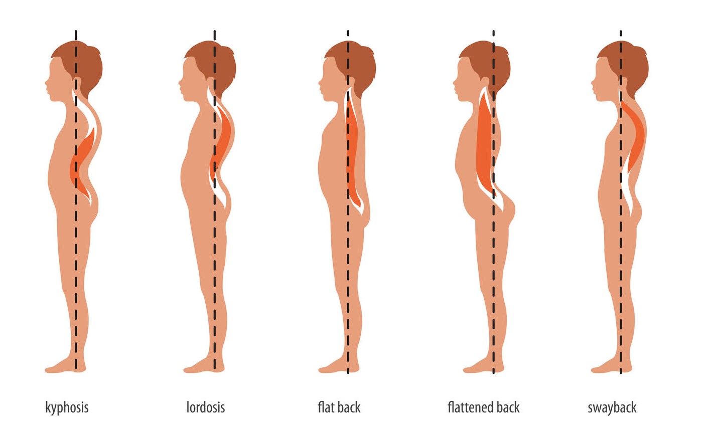
FAQs about Flatback Syndrome

MRI T2-weighted sagittal and axial images. Anterior kinking and... |  Download Scientific Diagram
MRI T2-weighted sagittal and axial images. Anterior kinking and... | Download Scientific Diagram

Visualizing misaligned cords with 2D sagittal flattening - Spinal Cord  Toolbox documentation
Visualizing misaligned cords with 2D sagittal flattening - Spinal Cord Toolbox documentation

A deep learning model for detection of cervical spinal cord compression in  MRI scans | Scientific Reports
A deep learning model for detection of cervical spinal cord compression in MRI scans | Scientific Reports

Automatic shape analysis. T2*WI of asymptomatic subjects showing... |  Download Scientific Diagram
Automatic shape analysis. T2*WI of asymptomatic subjects showing... | Download Scientific Diagram

Cervical Spondylotic Myelopathy (CSM) - Spinal Cord Compression - OrthoInfo  - AAOS
Cervical Spondylotic Myelopathy (CSM) - Spinal Cord Compression - OrthoInfo - AAOS

EPOS™
EPOS™

Neuroradiology threads by Lea Alhilali - Grading of Cervical Canal Stenosis  | Radiopaedia.org
Neuroradiology threads by Lea Alhilali - Grading of Cervical Canal Stenosis | Radiopaedia.org

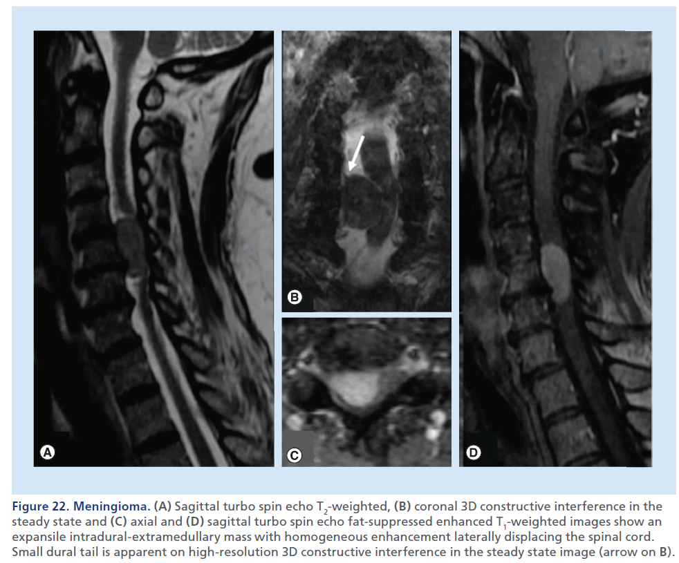
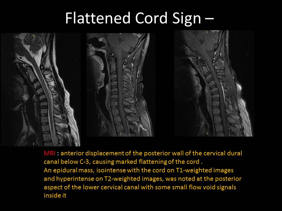
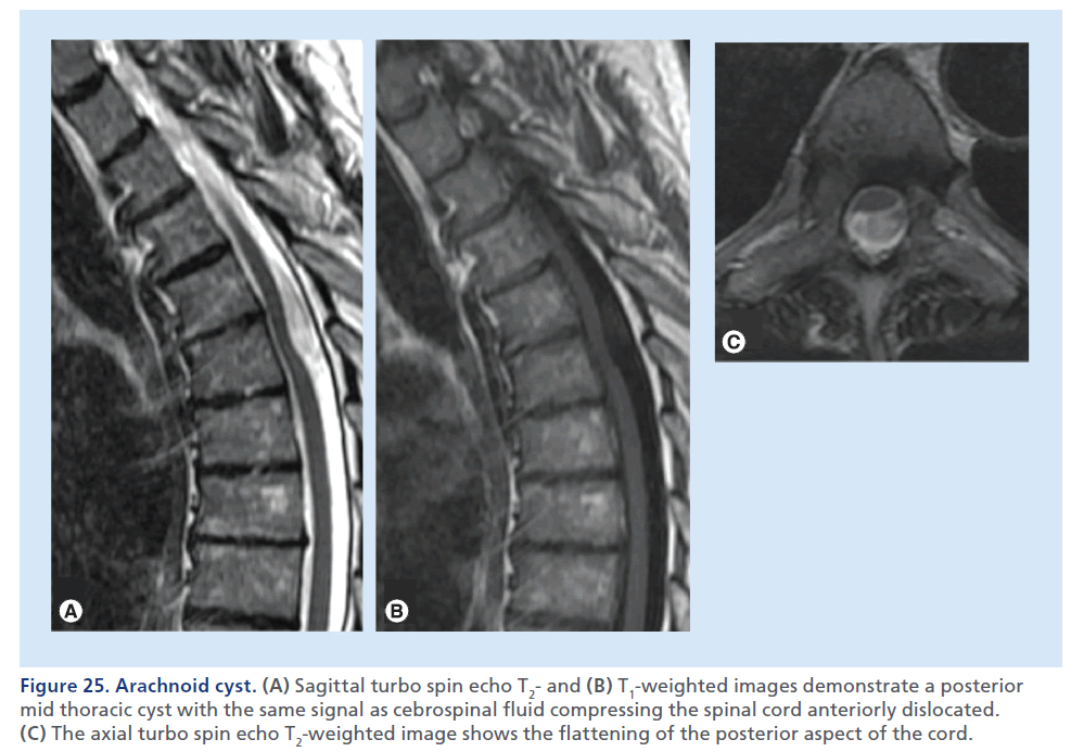
Imaging of spinal cord compression

Cervical Spondylotic Myelopathy (CSM) - Spinal Cord Compression - OrthoInfo  - AAOS
Cervical Spondylotic Myelopathy (CSM) - Spinal Cord Compression - OrthoInfo - AAOS

Nontraumatic Spinal Cord Compression: MRI Primer for Emergency Department  Radiologists | RadioGraphics
Nontraumatic Spinal Cord Compression: MRI Primer for Emergency Department Radiologists | RadioGraphics

Cauda Equina Syndrome - Complete Orthopedics | Multiple NY Locations

a Sagittal T2-weighted cervical MRI in neutral position shows marked... |  Download Scientific Diagram
a Sagittal T2-weighted cervical MRI in neutral position shows marked... | Download Scientific Diagram

A, The T2-weighted cervical magnetic resonance imaging (MRI) showed... |  Download Scientific Diagram
A, The T2-weighted cervical magnetic resonance imaging (MRI) showed... | Download Scientific Diagram

Lumbar lordosis flattens and the spinal canal adjusts around the static...  | Download Scientific Diagram
Lumbar lordosis flattens and the spinal canal adjusts around the static... | Download Scientific Diagram

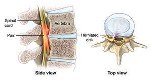
Spinal Cord Compression | Johns Hopkins Medicine

Dorsal Thoracic Arachnoid Web and the “Scalpel Sign”: A Distinct  Clinical-Radiologic Entity | American Journal of Neuroradiology
Dorsal Thoracic Arachnoid Web and the “Scalpel Sign”: A Distinct Clinical-Radiologic Entity | American Journal of Neuroradiology

Spinal Cord Compression | Johns Hopkins Medicine

A. T1 weighted MRI of the cervical spine demonstrates flattening and... |  Download Scientific Diagram
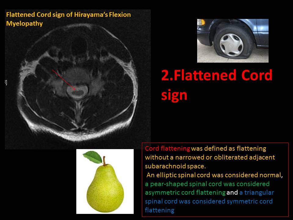
A. T1 weighted MRI of the cervical spine demonstrates flattening and... | Download Scientific Diagram

EPOS™
EPOS™

Imaging of spinal cord compression